Sep 29, 2023
Sanger Tree of Life HMW DNA Fragmentation: Covaris g-TUBE for ULI PacBio
- Graeme Oatley1,
- Filipa Sampaio1,
- Lucy Kitchin2,
- Raquel Juliana Vionette do Amaral1,
- Caroline Howard1
- 1Tree of Life, Wellcome Sanger Institute, Hinxton, Cambridgeshire, CB10 1SA;
- 2Wellcome Sanger Institute, Hinxton, Cambridgeshire, CB10 1SA
- Tree of Life at the Wellcome Sanger Institute
- Earth BioGenome Project


Protocol Citation: Graeme Oatley, Filipa Sampaio, Lucy Kitchin, Raquel Juliana Vionette do Amaral, Caroline Howard 2023. Sanger Tree of Life HMW DNA Fragmentation: Covaris g-TUBE for ULI PacBio. protocols.io https://dx.doi.org/10.17504/protocols.io.q26g7pm81gwz/v1
License: This is an open access protocol distributed under the terms of the Creative Commons Attribution License, which permits unrestricted use, distribution, and reproduction in any medium, provided the original author and source are credited
Protocol status: Working
We use this protocol and it's working
Created: September 11, 2023
Last Modified: September 29, 2023
Protocol Integer ID: 87631
Keywords: HMW DNA fragmentation, g-TUBE, ultra-low input, reference genome, long read sequencing, sanger tree of life hmw dna extraction protocol, life hmw dna extraction protocol, sanger tree of life hmw dna fragmentation, sanger tree of life hmw dna extraction, life hmw dna extraction, life hmw dna fragmentation, fragmentation of hmw dna, fragmented dna, sheared dna, dna extraction, hmw dna, effective for dna extract, dna extract, dna from sample, high molecular weight spri, abbreviations hmw, taxonomic groups in the tree, tree of life programme, extraction, dna, mediated fragmentation, taxonomic group, suitable for downstream long read, downstream long read
Funders Acknowledgements:
Wellcome Trust
Grant ID: 218328
Wellcome Trust
Grant ID: 206194
Gordon and Betty Moore Foundation
Grant ID: GBMF8897
Abstract
This protocol describes the centrifugation-mediated fragmentation of HMW DNA from samples prepared via any of the Sanger Tree of Life HMW DNA extraction protocols. The protocol produces fragments in the 8–10 kb size range using the Covaris g-Tube. The sheared DNA is suitable for downstream long read sequencing, including PacBio sequencing after ultra-low input (ULI) library preparation. This process is highly effective for DNA extracts across all taxonomic groups in the Tree of Life Programme. The output of this protocol is sheared DNA, which can be directed towards fragmented DNA clean up, using either the Manual or Automated SPRI protocols.
Acronyms and abbreviations
HMW: high molecular weight
SPRI: solid-phase reversible immobilisation
ULI: ultra-low input
Guidelines
- The DNA sheared using this protocol must be intended for ULI PacBio sequencing.
- This protocol is for the shearing of DNA into fragment sizes of 8–10 kb.
- 150 ng of unsheared DNA is ideal for this protocol, but 90–150 ng total input can be used.
Troubleshooting:
- Low concentration samples In some cases you may need to centrifuge >150 µL in order to process 150 ng. In this scenario, split your DNA sample into 100 µL or 150 µL subsamples, and centrifuge each subsample through the same g-TUBE. Re-combine the outputs from each centrifugation in preparation for downstream processes.
- Sample does not spin through the filter If, after three spins, the sample has not passed through the filter and still remains in the chamber, try the following:
- Flip the g-TUBE (screw cap down), and spin at 4,300 x g for 30 seconds.
- Recover the sample from the cap following the usual procedure (step 11).
- Filter the sample with a 0.45 µm filter.
- Use a new g-TUBE and follow the normal protocol.
Materials
- 1.5 mL DNA Lo-Bind microcentrifuge tubes (Eppendorf Cat. no. 0030 108.051)
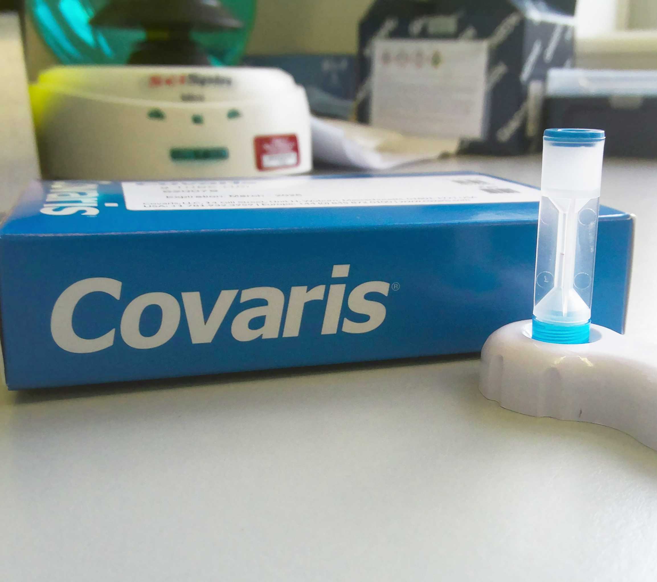
- Covaris g-TUBE with Covaris g-TUBE Prep Station (Cat. no. 520079)
- Buffer EB (Qiagen Cat. no. 19086)
Equipment
- Pipettes for 0.5 to 1000 μL and filtered tips
- Wide-bore tips (200 μL, filtered if available)
- Eppendorf™ Centrifuge 5425/5425 R (Cat. no. 5405000263)
Protocol PDF:  Sanger Tree of Life HMW DNA Fragmentation_ g-Tube for ULI PacBio.docx.pdf83.4KB
Safety warnings
- The operator must wear a lab coat, powder-free nitrile gloves and safety specs to perform the laboratory procedures in this protocol.
- Waste needs to be collected in a suitable container (e.g. plastic screw-top jar or Biobin) and disposed of in accordance with local regulations.
Laboratory protocol
Label the required number of Covaris g-TUBEs for each DNA sample that will be sheared; ensure that the tubes are labelled both on the lid and on the bottom.
Prior to transferring the DNA sample from its original tube, first mix the DNA sample by pipetting carefully with wide-bore pipette tip.
Transfer between 100–150 µL of the DNA sample to its corresponding labelled g-TUBE; 150 µL is the ideal volume.
If processing multiple samples, ensure that the volumes are equal by adding EB buffer to normalise the volumes. Be aware that the processing time from transfer of the sample into the g-TUBE and the g-TUBE containing the sample into the centrifuge should be no longer than 15 minutes, to avoid the sample migrating into the filter whilst on the bench.
Secure the g-TUBEs by tightly sealing the screw-caps, using the g-TUBE prep station provided with the g-TUBEs.
Place the g-TUBEs into a bench-top centrifuge. If there is an uneven number of samples, make sure to balance the centrifuge using a spare g-TUBE.
Run for 1 minute at 5,000 rpm for a shear size of 10 kb; refer to the manufacturer's instructions for conditions to obtain different shear sizes.
Repeat the centrifugation in step 7 until all the volume has passed through the filter, up to three times. Check the tube between chambers to ensure that all of your sample has passed through the filter. If the sample has not passed through the filter after the third spin, please refer to the ‘Troubleshooting’ section in the Guidelines section.
Invert the g-TUBEs within the prep station so the fixed base is facing up and the screw-cap is facing down, then repeat steps 6, 7 and 8 so that all of the DNA has passed completely through the g-TUBE filter a total of two times.
Once all of the sample has passed through the filter, remove the g-TUBEs from the centrifuge whilst maintaining their current orientation (screw-cap facing down), and place them screw-cap side down into the g-TUBE prep station.
Using the g-TUBE prep station to stabilise the tube, carefully unscrew the tube from the lid, leaving the lid in the prep station.
Aspirate the sample from the screw-cap using a standard pipette tip and transfer the sheared DNA to a fresh DNA Lo-Bind microcentrifuge tube.
Store the sheared DNA at 4 °C until further processing.
Protocol references