Sep 29, 2023
Sanger Tree of Life Fragmented DNA clean up: Manual SPRI

- Michelle Strickland1,
- Clare Cornwell1,
- Caroline Howard1
- 1Tree of Life, Wellcome Sanger Institute, Hinxton, Cambridgeshire, CB10 1SA
- Tree of Life at the Wellcome Sanger Institute
- Earth BioGenome Project

Protocol Citation: Michelle Strickland, Clare Cornwell, Caroline Howard 2023. Sanger Tree of Life Fragmented DNA clean up: Manual SPRI. protocols.io https://dx.doi.org/10.17504/protocols.io.kxygx3y1dg8j/v1
License: This is an open access protocol distributed under the terms of the Creative Commons Attribution License, which permits unrestricted use, distribution, and reproduction in any medium, provided the original author and source are credited
Protocol status: Working
We use this protocol and it's working
Created: September 04, 2023
Last Modified: September 29, 2023
Protocol Integer ID: 87308
Keywords: DNA clean up, solid phase reversible immobilisation, SPRI, manual SPRI, KingFisher, AMPure PB beads, reference genome, long read sequencing, sanger tree of life hmw dna fragmentation protocol, sanger tree of life hmw dna fragmentation, life hmw dna fragmentation protocol, sanger tree of life fragmented dna, life hmw dna fragmentation, hmw dna fragmentation, life fragmented dna, pacbio sequencing, fragmented dna, hmw dna, sheared dna, sheared dna from all of the taxonomic group, high molecular weight spri, using pacbio ampure pb bead, manual spri this protocol, read sequencing, pacbio ampure pb bead, manual spri, tree of life programme, spri, dna, sanger tree, pacbio, acronyms hmw, taxonomic group, including pacbio
Funders Acknowledgements:
Wellcome Trust
Grant ID: 218328
Wellcome Trust
Grant ID: 206194
Gordon and Betty Moore Foundation
Grant ID: GBMF8897
Abstract
This protocol describes the manual clean up of fragmented DNA following the Sanger Tree of Life HMW DNA Fragmentation protocols, using PacBio AMPure PB beads. This process is highly effective for the cleaning and removal of shorter fragments from sheared DNA from all of the taxonomic groups covered by the Tree of Life Programme. The output of this protocol is DNA which can be submitted for long read sequencing, including PacBio sequencing following Low Input (LI) or Ultra-Low Input (ULI) library preparation.
Acronyms
HMW: high molecular weight
SPRI: solid-phase reversible immobilisation
LI: low input
ULI: ultra-low input
Guidelines
- For DNA sheared using the Sanger Tree of Life HMW DNA Fragmentation: Diagenode Megaruptor® 3 for PacBio HiFi protocol, use a ratio of 0.6X AMPure PB beads to DNA volume.
- For DNA sheared using the Sanger Tree of Life HMW DNA Fragmentation: Diagenode Megaruptor® 3 for LI PacBio protocol, use a ratio of 1X AMPure PB beads to DNA volume.
- For DNA sheared using the Sanger Tree of Life HMW DNA Fragmentation: g-Tube for ULI PacBio protocol, use a ratio of 0.6X AMPure PB beads to DNA volume.
- To allow for QC to be performed and to meet internal requirements at Sanger for sequencing, 49 µL of EB is added in step 13 (3 µL is for QC and 45.4 µL for sequencing), however any volume of EB buffer can be used to elute the sheared DNA.
Materials
- 1.5 mL DNA Lo-Bind microcentrifuge tubes (Eppendorf Cat. no. 0030 108.051)
- AMPure beads PB (Pacific Biosciences Cat. no. 100-265-900)
- Buffer EB (Qiagen Cat. no. 19086)
- 100% absolute ethanol
- Nuclease-free water
- 15 mL or 50 mL centrifuge tubes
Equipment:
- Pipettes for 0.5 to 1000 μL and filtered tips
- Wide-bore tips (200 μL, filtered if available)
- DynaMag™-2 magnetic rack (Cat. no. 12321D) or similar
- Vortexer (Vortex Genie™ 2 SI-0266)
- Eppendorf ThermoMixer C (Cat. no. 5382000031) or similar
- Mini-centrifuge (Cat. no. SS-6050)
- Timer
Protocol PDF:  Sanger Tree of Life Fragmented DNA clean up_ Manual SPRI.docx.pdf69KB
Troubleshooting
Safety warnings
- The operator must wear a lab coat, powder-free nitrile gloves and safety specs to perform the laboratory procedures in this protocol.
- Waste needs to be collected in a suitable container (e.g. plastic screw-top jar or Biobin) and disposed of in accordance with local regulations.
- Liquid waste needs to be collected in a suitable container (e.g. glass screw-top jar) and disposed of in accordance with local regulations.
Before start
- AMPure PB beads are stored in the fridge at 4 °C – take them out 30 minutes before use to allow beads to equilibrate to room temperature.
- Set the heat block to 37 °C.
- Prepare fresh 80% ethanol. This solution is hygroscopic and should be prepared fresh each time to achieve optimal results using 100% absolute ethanol and nuclease free water.
- Prepare 3 labelled 1.5 mL microcentrifuge tubes for each sample to be used in steps 1, 9 and 17.
Laboratory protocol
Using a standard pipette tip, measure the post-shearing sample volume and transfer DNA solution from Diagenode tube to a new labelled 1.5 mL microcentrifuge tube. Record the volume.
Calculate the volume of AMPure PB beads needed for each sample based on the sheared DNA volume and the ratio required.
Vortex the AMPure PB beads for 30 seconds, then immediately add the calculated volume of beads to the sheared DNA sample.
Mix the bead/DNA solution thoroughly by pipette mixing 15 times with a wide-bore pipette tip. Do not flick the tube.
Quickly spin down the tube (for 1 second) on a mini-centrifuge to collect the beads.
Incubate the mix on the bench top for 5 minutes at room temperature.
Spin down the tube (for 1 second) on a mini-centrifuge to collect the beads.
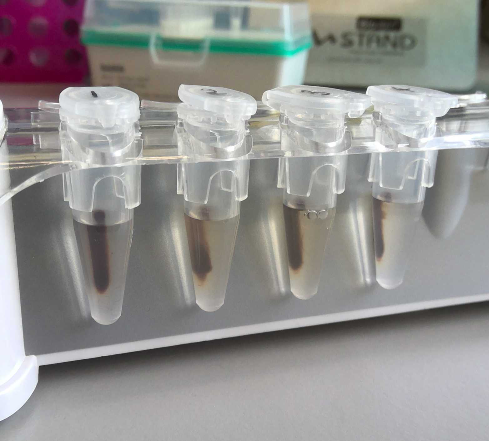
Place the tube in a magnetic bead rack and wait for the beads to pellet on the side of the tube. This can take approximately 1-5 minutes.
Slowly pipette off the cleared supernatant from the tube and save (in another labelled 1.5 mL microcentrifuge tube). Avoid disturbing the beads.
Wash the pelleted beads with freshly prepared 80% ethanol.
- Do not remove the tube from the magnetic rack.
- Use a sufficient volume of 80% ethanol to fill the tube (1.5 mL for 1.5 mL microcentrifuge tube).
- Slowly dispense the 80% ethanol against the side of the tube opposite the beads, taking care not to disturb the pelleted beads.
- After 30 seconds, pipette and discard the 80% ethanol.
Repeat step 10.
Spin down tubes on a mini-centrifuge for 1 second and return them to the magnetic rack, allowing the beads to pellet. Aspirate and dispose of any remaining ethanol.
Check for any remaining ethanol droplets in the tube. If droplets are present, repeat step 12.
Take tubes off the magnetic rack and add 49 µL of EB buffer to the beads. Gently mix by slowly pipetting 15 times with a wide-bore pipette tip. Do not flick the tube.
Incubate tubes at 37 °C for 15 minutes.
Briefly spin down the tubes (for 1 second) on a mini-centrifuge and place them in the magnetic rack. Allow beads to pellet.
Without disturbing the beads, transfer the supernatant to a new 1.5 mL DNA Lo-Bind microcentrifuge tube.
Perform QC as required.
Store samples at 4 °C.
Protocol references
