Feb 23, 2018
Version 2
Human Liver Caudate Lobe Dissociation for ScRNA-seq V.2
- Sonya MacParland1,
- Xue-Zhong Ma1,
- Justin Manuel1,
- Jeff Liu1,
- Gary Bader1,
- Ian McGilvray1
- 1University of Toronto
- Human Cell Atlas Method Development Community


Protocol Citation: Sonya MacParland, Xue-Zhong Ma, Justin Manuel, Jeff Liu, Gary Bader, Ian McGilvray 2018. Human Liver Caudate Lobe Dissociation for ScRNA-seq. protocols.io https://dx.doi.org/10.17504/protocols.io.m9sc96e
License: This is an open access protocol distributed under the terms of the Creative Commons Attribution License, which permits unrestricted use, distribution, and reproduction in any medium, provided the original author and source are credited
Protocol status: Working
We use this protocol and it's working
Created: February 14, 2018
Last Modified: March 28, 2018
Protocol Integer ID: 10258
Keywords: human liver caudate lobe dissociation for scrna, viable resident cells from liver sample, human liver caudate lobe dissociation, cell dissociation, cell dissociation method, process of cell dissociation, deceased donor liver graft, liver sample, human liver, scrna, organ transplantation, damaging to the cell, viable resident cell, single cell level, transcriptome, biasing of the transcriptome, cell
Abstract
One of the challenges to describing the human liver on a single cell level is the process of cell dissociation. Cell dissociation methods can be quite damaging to the cells and can lead to a biasing of the transcriptome. We have developed gentle surgical and perfusion techniques to isolate and transcriptionally profile viable resident cells from liver samples taken from deceased donor liver grafts prior to organ transplantation.
Troubleshooting
Before start
Prior to collagenase and neutral protease digestion, solution 3 should be warmed to 37 degree celsius.
Before starting any perfusions, all solutions must be oxygenated with 95%Os2 and 5% CO2.
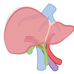
Surgical Resection of Human Liver Caudate Lobe
Resection of the human liver caudate lobe (segment 1) is carried out on the surgical backbench as part of the preparation of the organ for implantation. The liver is flushed at the donor hospital with University of Wisconsin (UW) solution or Histidine-tryptophan-ketoglutarate (HTK) solution (Methapharm).
UW solution
https://www.ncbi.nlm.nih.gov/pubmed/1689516
00:10:00
Transport of Human Liver Caudate Lobe for Cell Isolation
Transport of caudate lobe in cold histidine tryptophan−ketoglutarate (HTK) solution (Methapharm) to cell isolation suite.
00:15:00
Cannulation of the Human Liver Caudate Lobe
Cannulation of caudate lobe with two or three irrigation cannulae with olive tips inserted into exposed vessels in the cut surface of the liver lobe. Once placed, cannulae are secured with surgical glue (3M Vetbond: 1469SB).
Vendor for cannulae: http://www.acufirm.de/index.php?seite=119
Manufacturer: Ernst Kratz GmbH
Catalogue number for irrigation cannulae: 1464LL (straight cannulae, 1.2mm or 2mm in diameter), 1465LL (curved cannulae, 1.2mm or 2mm in diameter).
Note
Irrigation cannulae (which differ in diameter and whether they are curved or straight) are selected based on the size of caudate and the size and location of the exposed vasculature.
CRITCAL POINT: Following cannulation and during all steps of the perfusion, the caudate should be wrapped in moist gauze (moistened with the perfusion media) to prevent the drying of the Glisson's capsule and the exposed tissue.
Human Liver Caudate Lobe Perfusion step 1
Caudate perfusion with Hank’s balanced salt solution (HBSS)+EGTA 10mM for 15-20 min. Perfusion performed at 4 degrees celsius. Perfusion rate is 10mL/min/cannulae. Perfusion is carried out using a peristaltic pump (MasterFlex L/S- Cole Palmer).
| Solution 1 | Sodium Choride | 152.5mM | |||
| HEPS | 19.8mM | ||||
| Potassium Chloride | 5.5mM | ||||
| Glucose (D-glucose) | 5.0mM | ||||
| Sodium hydrogen carbonate (NaHCO3) | 24.8mM | ||||
| EGTA | 0.1mM |
Note
All solutions are oxygenated with 95%O2 and 5% CO2 prior to perfusion
00:15:00
Note
CRITICAL POINT: Duration of perfusion based on the size of the caudate and the completeness of the perfusion.
Human Liver Caudate Lobe Perfusion step 2
Caudate perfusion with HBSS + CaCl2 dehydrate 0.5µM for 15-20min. Perfusion performed at 4 degrees celsius. Perfusion rate is 10mL/min/cannulae.
| Solution 2 | Sodium Choride | 152.5mM | |||
| HEPS | 19.8mM | ||||
| Potassium Chloride | 5.5mM | ||||
| Glucose (D-glucose) | 5.0mM | ||||
| Sodium hydrogen carbonate (NaHCO3) | 24.8mM | ||||
| Calcium Chloride Dihydrate | 0.5uM |
All solutions are oxygenated with 95%O2 and 5% CO2 prior to perfusion.
Duration of perfusion based on the size of the caudate and the completeness of the perfusion.
00:20:00
Collagenase/Neutral Protease Digestion
Digestion is carried out by perfusion with collagenase (Collagenase MA; Vitacyte Cat#001-2030) plus neutral protease (BP Protease; VitaCyte Cat#003-1000) in solution 3, according to the manufacturers’ suggested protocol. The concentration of collagenase used is dependent on the collagen degradation activity (CDA) of each collagenase batch. The concentation of neutral protease employed is based on the neutral protease acitivity (NPA) for each batch. Each caudate lobe is perfused with 0.25 Million CDA units/Caudate and 0.25 Million NP units per caudate in 100mL of Solution 3. The volume used to perfuse may be increased if the caudate is larger than 30 grams. Digestion is carried out at 37 degrees celsius. Perfusion rate is 10mL/min/cannulae.
CRITICAL POINT: Perfusion is carried out in a recirculation manner for 15 to 20 minutes or until the liver appeared to break apart slightly under Glisson’s capsule. Two MasterFlex L/S (Cole-Palmer) pumps are employed for the recirculation, One delivering perfusate to the caudate (in-flow) and 1 collecting and returning the outflow to the oxygenated perfusion solution.
| Solution 3 | Sodium Choride | 152.5mM | |||
| HEPS | 19.8mM | ||||
| Potassium Chloride | 5.5mM | ||||
| Glucose (D-glucose) | 5.0mM | ||||
| Sodium hydrogen carbonate (NaHCO3) | 24.8mM | ||||
| Calcium Chloride Dihydrate | 4mM |
Calcium Chloride Dihydrate concentration is increased compared to solution 2 as calcium ions are required for collagenase enzyme stability and activity.
All solutions are oxygenated with 95%O2 and 5% CO2 prior to perfusion.
Duration of perfusion based on the size of the caudate and the completeness of the perfusion.
00:20:00
Dissociation
The digested lobe is placed on a crystallizing dish containing 100-200ml of HBSS with 0.1% human albumin (Sigma) (human albumin can be substituted with 10% fetal calf serum to inactivate the collagenase and the neutral protease).
A scalpel is used to cut through the tissue and release cells contained within. The remaining tissue should be gently agitated by hand with tweezers to release the cells, employing minimal or no mechanical dissociation.
00:05:00
Preparation of Liver Single Cell Suspension for ScRNA-seq
Liver homogenate is filtered through a 70uM filter (ThermoFisher Scientific; Cat. No. 08-771-2).
The lobe is flushed at the donor hospital so no red cell lysis step is required (if adapting this protocol for core or fine needle aspiration biopsies, red blood cell lysis should be employed if the sample appears red or pink tinged).
We do not include flow cytometry-based, density gradient-based or column purification-based steps, whether to enrich for cell populations or to remove dead or dying cells. We avoid these steps as sample manipulation can lead to further losses in cells. Low quality cells with low library sizes are removed during the data analysis steps.
00:05:00
Count and assess viability by trypan blue.