Oct 21, 2024
Version 2
FUNDIS Fungal Tissue Sampling for PCR V.2
- Harte Singer1,2,3,4
- 1FUNDIS;
- 2Dikarya LLC;
- 3Cal State East Bay;
- 4Entheome Foundation

Protocol Citation: Harte Singer 2024. FUNDIS Fungal Tissue Sampling for PCR. protocols.io https://dx.doi.org/10.17504/protocols.io.eq2lyw93rvx9/v2Version created by Harte Singer
License: This is an open access protocol distributed under the terms of the Creative Commons Attribution License, which permits unrestricted use, distribution, and reproduction in any medium, provided the original author and source are credited
Protocol status: Working
We use this protocol and it's working
Created: July 19, 2024
Last Modified: October 21, 2024
Protocol Integer ID: 110348
Abstract
How to sample dried fungi into PCR tubes for DNA extraction.
Materials
- Your mushrooms - kept together in a box or a bag.
- Another box or bag to place finished specimens
- Tweezers -a fine tip non-serrated for most specimens and a dissecting needle for any tough material.
- A fine-tip Sharpie
- A box of Kimwipe tissues
- A small glass or plastic container with some 70% isopropyl rubbing alcohol.
- 12 strips of 8-well PCR tubes in a PCR tube rack
- An empty PCR tube rack
- A trash vessel to discard dirty kimwipes.
- A device to access your data entry spreadsheet
- (Optional) A pair of nitrile gloves
- (Optional) Razor Blade/Scalpel
- (Optional) A pencil
- (Optional) A printout of the instructions for reference
- (Optional) A printout of your 96 tube rack template
Sampling guide
Sampling guide
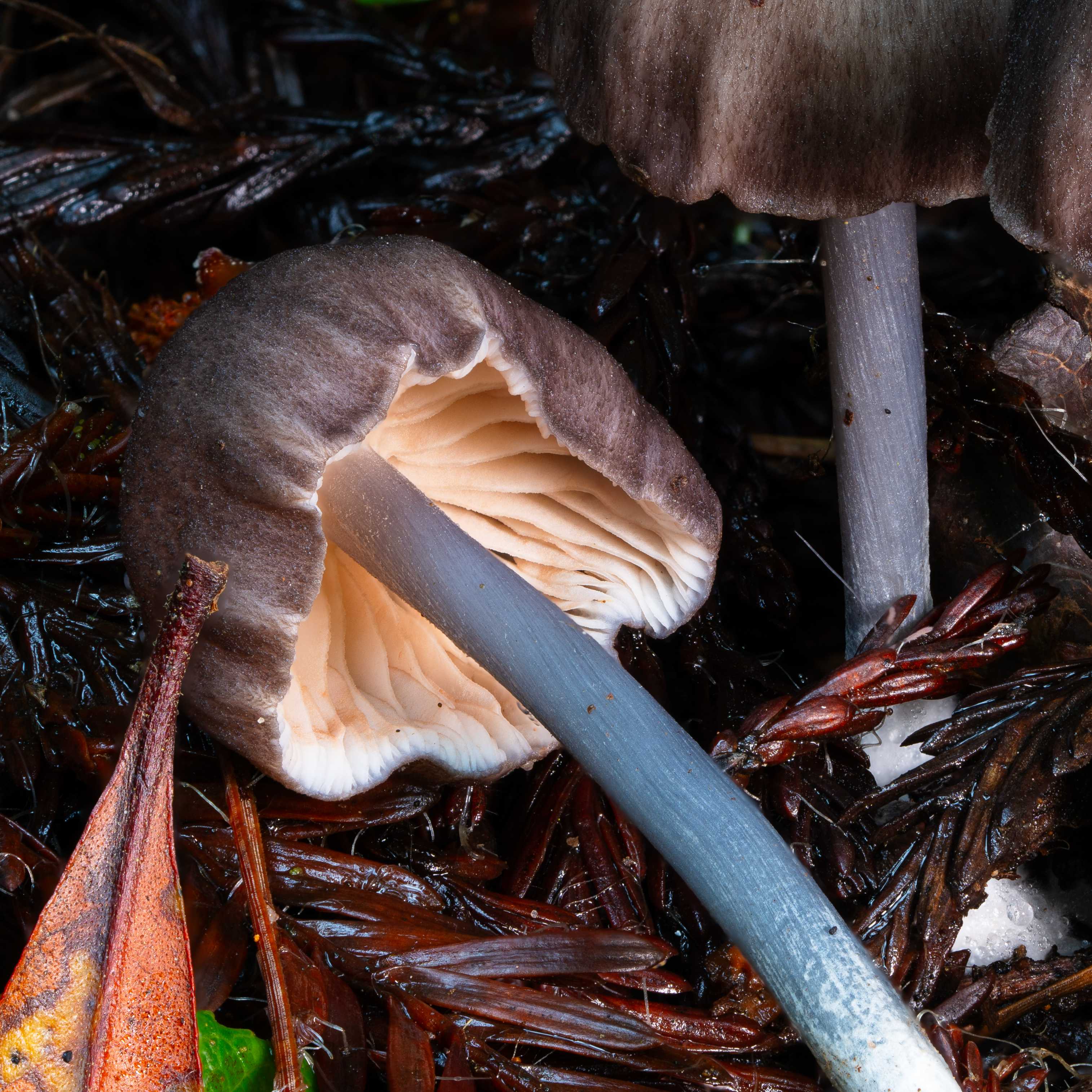
Gilled Mushrooms - Sample a very small piece of the gill.
Mushrooms with pores - Sample a very small piece from the pore tissue
Cup fungi - Sample from the inner surface of the cup - the part that generally faces away from the substrate. Sometimes there will be a stalk and sometimes there isn't.
Inverted Cup Fungi - Sample from the exterior of the tissue on the end of the stalk
Truffles - Sample from the interior of the mushroom. Typically there will be one or more samples that are sliced in half.
Puffballs - These are similar to truffles in appearance, but contain a soft, often powdery interior. Sample the inner material, but be very careful to avoid releasing a puff of spores. It is a good idea to save these for last to avoid cross contamination. Avoid breathing spores.
Crusts - These are mushrooms that are flat against a substrate. They can have many textures such as pores, teeth, or they can be smooth. In general, you can sample any part of these, but be careful not to dig into the substrate. Sample by scraping a bit of material off with the tweezers if the surface is completely flat.
Bird's Nests - These are little cups that contain "eggs" called peridioles. Try to break apart a peridiole and sample that material.
Tissue Sampling
Tissue Sampling
2h
2h
Find a suitable place to conduct tissue sampling. A hard, nonporous surface that can be wiped clean with rubbing alcohol is ideal. Turn off any fans and close any windows so there is minimal air movement. Wash your hands and gather your materials and mushrooms for sampling.
Set up one of the PCR tube racks and with very clean hands, remove each strip of PCR tubes from its package and close each of the lids before placing the strip into a row of the empty PCR tube rack.
Using a sharpie, label the hinges of each PCR strip using the pattern depicted in the photo. Also label the first tube in each strip from 1-12 going from the top to bottom as depicted. Additionally, you can label the last tube in each strip with the plate number, so if this was plate number 5, the last tube on the right of each strip would be labeled 5. This is not depicted in this photo.
Dip your tweezers into the alcohol and then wipe them using a piece of clean kimwipe. You don't need a whole square and can just rip off a small piece, but make sure to wipe the tweezers thoroughly and repeat this until you are confident they are clean. Alcohol will not destroy DNA, it just helps remove the debris on the tweezer. Most mushrooms don’t make a ton of debris, but be extremely careful with puffballs or any mushroom with a lot of powdery spores that tend to become airborne. It is best to save these specimens for the end of sampling to avoid potentially cross-contaminating
Set tweezers down such that the tines do not come in contact with anything while you grab the mushroom.
Remove the lid from the tube rack, then remove the first strip of 8 tubes from the tube rack and place it in the second rack for sampling. The first tube on the left will have 1 written on it and the first hinge will be marked.
Grab a box/bag of mushrooms and enter the accession number into the data entry sheet in the first corresponding cell. Write the tube number back side of the voucher slip (i.e. 1-1, 1-2 … 12-8).
Open the box/bag of mushrooms and either reach in with the tweezer or take out the mushroom and place on a clean surface away from your tubes. Carefully remove a small piece of tissue. You will only need a piece about 1x1mm. Hymenium tissue works best. Gills, pores, gleba, apothecia etc. This tissue has a high density of DNA. It is critical that you only take clean tissue. Any dirt, plant matter, or other contaminants will negatively impact the results. Take care not to cause too much damage to the specimens. Just take your time and take as little as you can. Too much tissue will negatively impact the results. Make sure you do this operation away from your tube rack to minimize the chance of cross contamination. The amount depicted in these photos is about the maximum amount of tissue you want to use
Place the tissue into the open tube and release it, or gently tap/wipe the tines inside the tube. Remove the tweezer and place it aside to be cleaned and close the cap. Static electricity can make samples stick to the sides of the tubes or make the bits of mushroom behave in a way you dont expect. Be mindful in your movements.
Clean your tweezers and proceed with the next specimen.
Place each completed strip back in its row in the original tube rack and remove the next strip of tubes for sampling.
Place the plastic cover on the tube rack and use lab tape or masking tape to tape the lid shut. Label the base of the tube rack and the lid using lab tape or masking tape.
Pack your tube rack in a box with plenty of padding for best shipping conditions.
